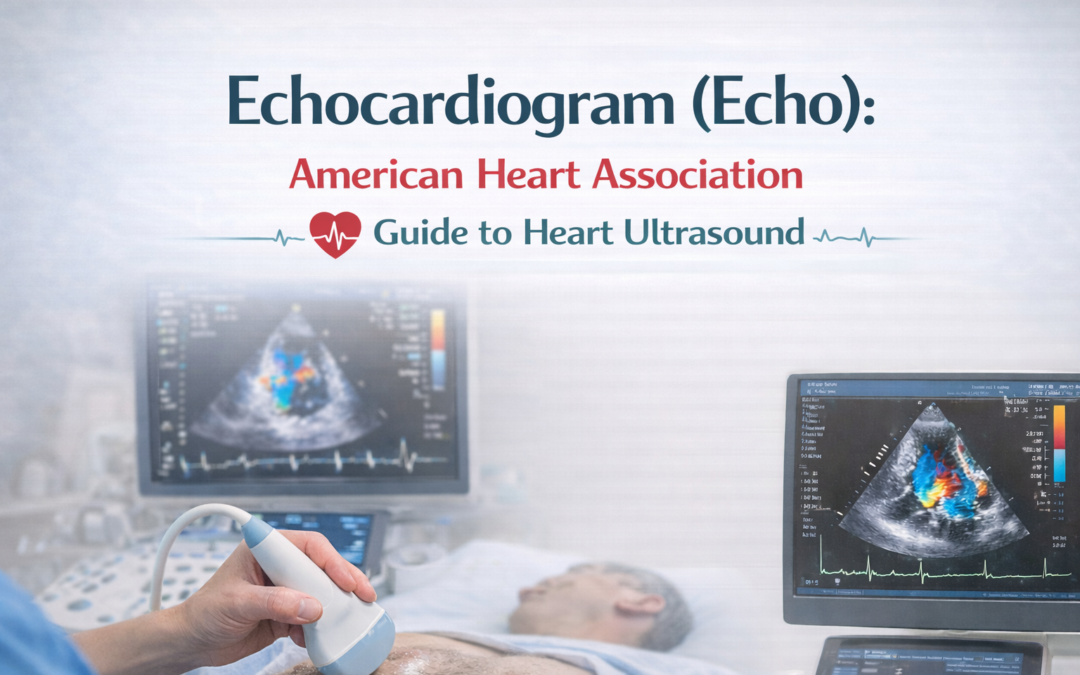
Overview of an Echocardiogram (Echo)
An echocardiogram, often called an echo, is a noninvasive ultrasound test that uses sound waves to create real-time images of the heart. It allows doctors to assess heart structure, blood flow, and overall cardiac function without radiation, making it a safe and commonly used diagnostic tool.
This test is frequently recommended to evaluate symptoms such as chest pain, shortness of breath, irregular heartbeat, or suspected heart disease. Echocardiography provides detailed information about heart chamber size, wall motion, valve structure, and pumping efficiency. It plays a key role in diagnosing heart conditions, monitoring existing disease, and guiding treatment decisions, especially in patients at risk for heart failure.
How Does Echocardiography Work?
During the test, a trained sonographer places a handheld device called a transducer on the chest. The transducer emits high-frequency sound waves that reflect off heart structures. These returning echoes are converted by a computer into moving images of the heart.
Advanced techniques such as Doppler and color flow imaging measure blood flow direction and speed, helping identify abnormalities.
Key imaging modes include:
- Two-dimensional (2D) imaging: Shows heart structure and movement
- M-mode: Measures chamber size and wall motion
- Doppler imaging: Assesses blood flow and velocity
- Color flow imaging: Visualizes blood flow patterns across valves and chambers
What Are the Risks of an Echocardiogram?
A transthoracic echocardiogram (TTE) is very safe and carries minimal risk. It does not use ionizing radiation and is routinely performed in cardiology settings.
A stress echocardiogram may rarely cause symptoms such as chest discomfort, irregular heartbeat, or shortness of breath during exercise or medication-induced stress. Medical staff closely monitor patients throughout the test.
A transesophageal echocardiogram (TEE) involves sedation and passing a probe into the esophagus. Possible risks include sore throat, nausea, or, very rarely, esophageal injury. Serious complications are uncommon.
What Can I Expect After the Test?
After a standard transthoracic echo, most people can return to normal activities immediately.
Most patients resume normal activities the same day without restrictions.
Following a stress echo, you may be observed briefly until heart rate and blood pressure return to normal. After a transesophageal echo, you will need to wait until sedation wears off and your gag reflex returns before eating or drinking.
Your healthcare provider will review the results and discuss any follow-up testing or treatment if needed.
When Should I Call My Doctor?
Contact your doctor if you experience chest pain, fainting, persistent dizziness, difficulty swallowing, or shortness of breath after the test. Seek urgent care if chest pain is severe or prolonged, especially after a stress echocardiogram.
Food and Medications Before an Echocardiogram
For a routine transthoracic echo, no special preparation is usually required. You may eat and take medications as prescribed.
Before a stress echocardiogram, you may be asked to avoid food, caffeine, or certain medications for several hours. For a transesophageal echo, fasting is required due to sedation. Always follow instructions provided by your healthcare team.
How the Test Will Feel
A transthoracic echocardiogram is painless. Gel is applied to the chest, and the transducer may press firmly but should not cause discomfort.
During a stress echo, you may feel tired or short of breath as your heart works harder. A transesophageal echo requires throat numbing and sedation, and mild throat soreness afterward is common.
What Abnormal Results May Indicate
Abnormal echocardiogram findings may include reduced pumping function, enlarged heart chambers, abnormal wall motion, valve disease, fluid around the heart, or increased pressure in the lungs. Results are interpreted in context with symptoms and medical history and may lead to further testing or treatment.
Preparing for Your Echocardiogram
Wear comfortable clothing and avoid applying lotions or oils to your chest on the day of the test. Bring a list of current medications. If you are scheduled for a stress or transesophageal echo, follow fasting and medication instructions carefully and arrange transportation if sedation is used.
After Your Echocardiogram
Most patients resume normal activities immediately after a transthoracic echocardiogram. After a stress or transesophageal echo, short-term monitoring may be required. Avoid driving or operating machinery for 24 hours if sedated.
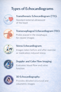
Types of Echocardiograms
- Transthoracic Echocardiogram (TTE): Standard external ultrasound of the heart
- Transesophageal Echocardiogram (TEE): Probe placed in the esophagus for clearer images
- Stress Echocardiogram: Images taken before and after exercise or medication-induced stress
- Doppler and Color Flow Imaging: Evaluates blood flow and valve function
- 3D Echocardiography: Provides detailed structural and volumetric images
Frequently Asked Questions
1. What should you avoid before an echocardiogram?
For a routine transthoracic echo, avoid heavy chest creams. For stress or transesophageal echocardiograms, follow fasting and medication instructions and avoid caffeine if advised.
2. What can an echocardiogram detect?
It can detect heart chamber enlargement, reduced heart function, valve abnormalities, congenital defects, fluid around the heart, and blood clots.
3. How long does an echocardiogram take?
A standard transthoracic echocardiogram usually takes 30–60 minutes. Stress and transesophageal echocardiograms may take longer due to preparation and recovery time.
4. Are ECG and echocardiogram the same?
No. An ECG records the heart’s electrical activity, while an echocardiogram uses ultrasound to visualize heart structure and movement. They provide complementary information.
5. What are the benefits and risks of an echocardiogram?
Benefits include noninvasive, radiation-free imaging that helps diagnose and manage heart conditions. Risks are minimal, with rare complications in stress or transesophageal studies.